Illustrations of Histology
Lesson 3
Nervous System
Objectives
By the end of the video, students should be able to:
Describe the structure of the central nervous system (brain and spinal cord), the location of white and gray matter, and the function for the observed gross structure including the sulci, gyri, and location of the white and gray matter.
Name the structure and function of the key glial cells within the central nervous system including astrocytes, oligodendrocytes, microglia, and ependymal cells.
Name all of the locations where cerebrospinal fluid fills the ventricles and canals within the CNS.
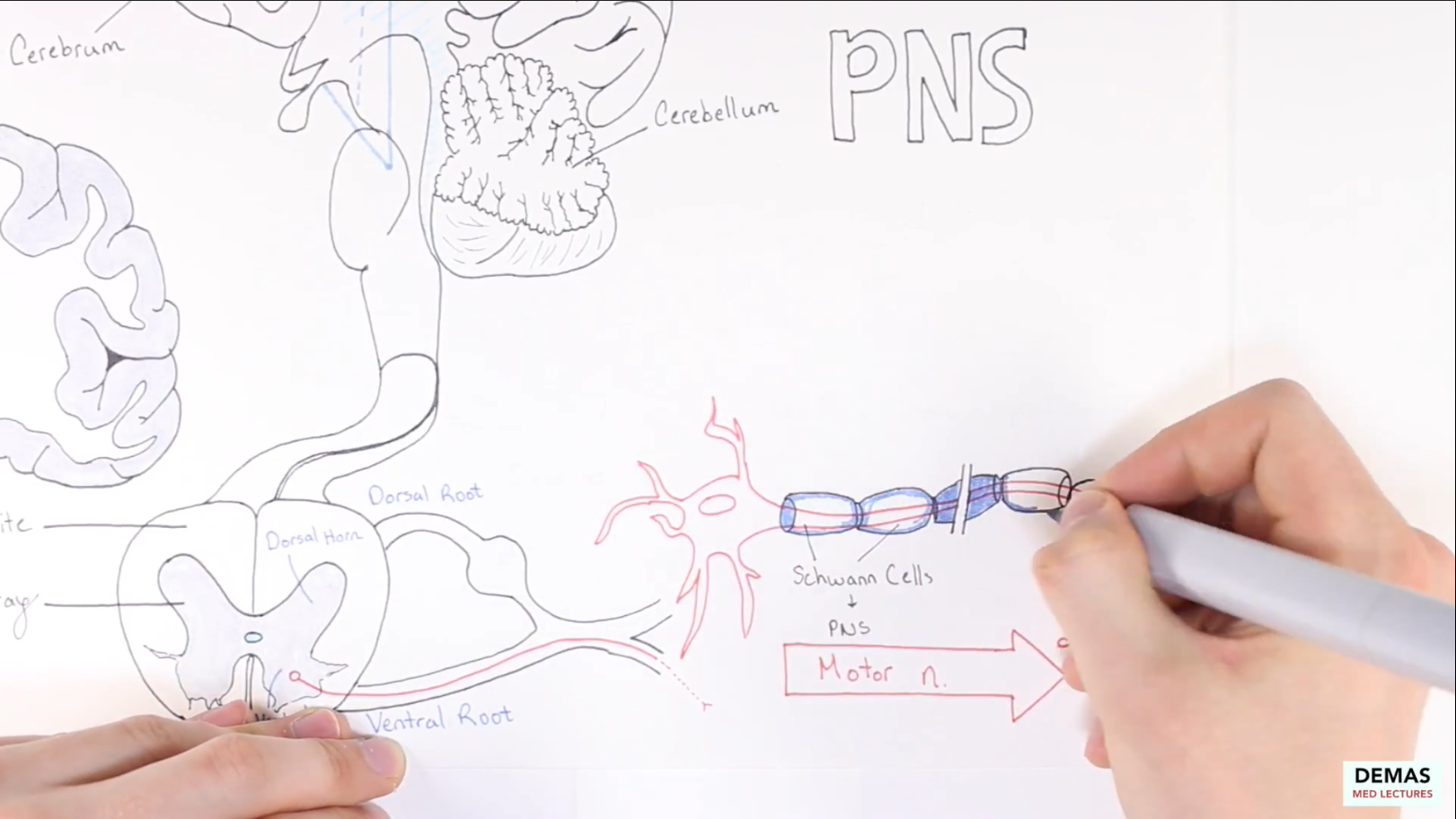
Name and label all of the important structures of the motor and sensory neuron within the PNS including the cell body, axon, axon terminal, schwann cells, and myelin sheath.
Recall the number of layers of the cerebral cortex and cerebellum and be able to label the structures of the cerebellum only as seen on H&E stained slides.
To Do List:
Watch lecture video
Download and review handout
Complete post-lecture quiz
Lecture 3
Handouts
Review handout can be downloaded here
