Illustrations of Histology:
Lesson 4
Blood cells
Objectives
By the end of the video, students should be able to:
Identify the composition and abundance of the three distinct layers of blood as it would appear when separated within an anticoagulated test tube.
Describe the key features of red blood cells (shape and cellular components) and how this contributes to their function
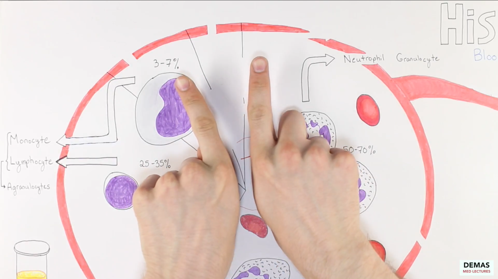
Describe the key elements of each of the white blood cells: relative size/abundance, their characteristic appearance on Wright Giemsa stain, function, and whether they are considered an agranulocyte or granulocyte.
Understand which cells are found within the blood vessel and the cells that are found only outside of the vasculature in the mature form.
To Do List:
Watch lecture video
Download and review handout
Complete post-lecture quiz
Lecture 4
Handouts
Review handout can be downloaded here
